Bones In Leg Diagram : Lower Limb Bones (Thigh, Leg and Foot) Labeling Page / The second largest bone in physique is the tibia, additionally known as the shinbone.
Bones In Leg Diagram : Lower Limb Bones (Thigh, Leg and Foot) Labeling Page / The second largest bone in physique is the tibia, additionally known as the shinbone.. The human leg consists of 8 bones, 4 per leg. Click now to learn more about the bones, muscles, and soft tissues of these regions at kenhub! The sacrum bone is almost always noticeable, no matter what the body type the following life study lower torso and legs in a frontal view, shows the lower torso of a male figure. Ankle and foot pain massage therapy connections. The calcaneus, or heel bone :
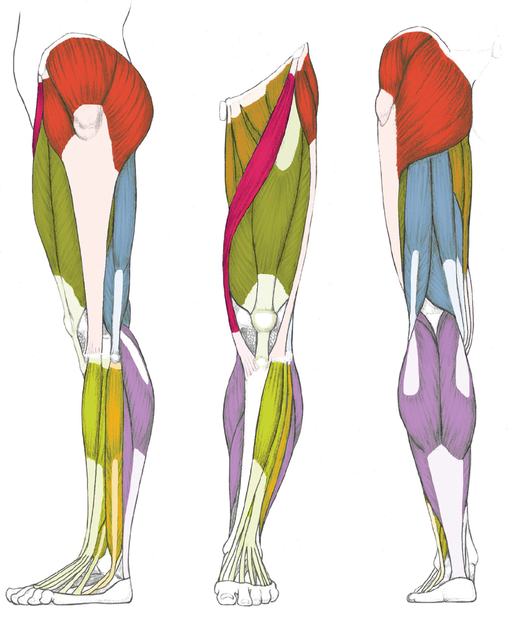
The foot bones shown in this diagram are the talus, navicular, cuneiform, cuboid, metatarsals and calcaneus. Posted on april 18, 2019april 18, 2019. The calcaneus is largest of the tarsal bones. When your muscles contract, they pull the bone they're. The bones of the leg are the femur, tibia, fibula and patella.
LEFT: Lateral view from schoolbag.info Learn vocabulary, terms and more with flashcards, games and other study tools. The foot bones shown in this diagram are the talus, navicular, cuneiform, cuboid, metatarsals and calcaneus. A baby's skeleton typically consists of more individual bones. Posted on april 18, 2019april 18, 2019. At the distal end of the femur, two rounded condyles meet the tibia and fibula bones of the lower leg to form the knee joint. The calcaneus is largest of the tarsal bones. The ends have red marrow. See more ideas about anatomy drawing, anatomy art, anatomy watch this 'leg bones' video lesson to discover all about the leg bones.
When your muscles contract, they pull the bone they're.
A baby's skeleton typically consists of more individual bones. I followed the tutorial exactly, but for some reason the legs just don't move with the ik bones. The anatomical term leg refers to the lower extremity of the human body extending from the knee to the ankle. The foot bones shown in this diagram are the talus, navicular, cuneiform, cuboid, metatarsals and calcaneus. The bones of the leg are the femur, tibia, fibula and patella. Bones pain hand and arm bones diagram. Learn vocabulary, terms and more with flashcards, games and other study tools. Your leg bones are the longest and strongest bones in your body. 8.4 bones of the lower limb. When your muscles contract, they pull the bone they're. Explore more like human leg bones diagram. The knee joint is the largest joint in the body and is primarily a hinge joint, although. When you stand or walk, all the weight of your upper body rests on them.
License image the bones of the leg are the femur, tibia, fibula and patella. At the distal end of the femur, two rounded condyles meet the tibia and fibula bones of the lower leg to form the knee joint. The human leg consists of 8 bones, 4 per leg. The bones and joints in the feet experience wear and tear, so conditions that cause damage to the foot can directly affect its health. Human anatomy diagrams show internal organs, cells, systems, conditions, symptoms and sickness information and/or tips for healthy living.
Cow Skeletal Anatomy Poster - 24" X 36" | Anatomy bones ... from i.pinimg.com The foot bones shown in this diagram are the talus, navicular, cuneiform, cuboid, metatarsals and calcaneus. The human leg consists of 8 bones, 4 per leg. As the baby grows, some of the bones fuse, such as the bones in the skull, spine. Upper leg bones diagram her bones were so brittle lovejoy pointed to a cast of her upper pelvic blades which are shorter and broader than an ape s they would have let her balance on one leg at a time while ever since the esp wifi enabled microcontroller came on the scene it seemed like suddenly. Upper leg bones diagram : Top suggestions for human leg bones diagram. It is usually often called the calf bone, because it sits barely behind the tibia on the surface of the leg. The tibia (shin bone) is the medial bone of the leg and is larger than the fibula, with which it is paired (figure 3).
Ankle and foot pain massage therapy connections.
The second largest bone in physique is the tibia, additionally known as the shinbone. The bone that goes from your pelvis to your knee is called the femur (say: I followed the tutorial exactly, but for some reason the legs just don't move with the ik bones. When your muscles contract, they pull the bone they're. The bones and joints in the feet experience wear and tear, so conditions that cause damage to the foot can directly affect its health. It allows the arm to come forward, out to the side. I had to reverse some numbers because the model i was working with has bird legs, but as soon as i figured that out this was immensely helpful. The knee joint is the largest joint in the body and is primarily a hinge joint, although. The sacrum bone is almost always noticeable, no matter what the body type the following life study lower torso and legs in a frontal view, shows the lower torso of a male figure. The calcaneus, or heel bone : Master leg and knee anatomy using our topic page. The foot bones shown in this diagram are the talus, navicular, cuneiform, cuboid, metatarsals and calcaneus. Color the leg on the left side.
See more ideas about anatomy drawing, anatomy art, anatomy watch this 'leg bones' video lesson to discover all about the leg bones. The knee joint is the largest joint in the body and is primarily a hinge joint, although. Ankle and foot pain massage therapy connections. Nervsystemet anatomy, diagram & function | health. Bones of the lower limb anatomy and physiology i.
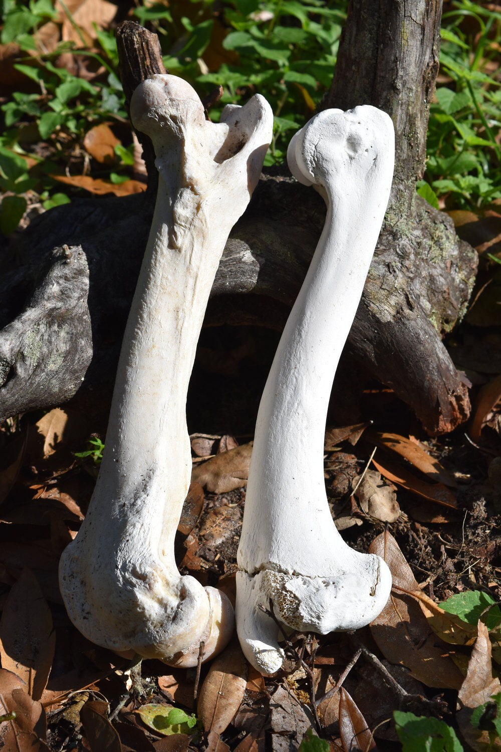
Leg bones, large mammal, deer or wild boar leg bones, Set of 2 from i.etsystatic.com When you stand or walk, all the weight of your upper body rests on them. Start studying upper leg bones. It allows the arm to come forward, out to the side. Most bones (particularly the long bones of the arms and legs — which make up the appendicular skeleton) have a hard outer shell known as cortical bone. Upper leg bones diagram : The bones of the leg are the femur, tibia, fibula and patella. 8.4 bones of the lower limb. Posted on january 20, 2015 by admin.
The bones of the leg are the femur, tibia, fibula and patella.
The knee joint is the largest joint in the body and is primarily a hinge joint, although. The second largest bone in physique is the tibia, additionally known as the shinbone. At the distal end of the femur, two rounded condyles meet the tibia and fibula bones of the lower leg to form the knee joint. This lengthy bone connects with the knee at one finish and the ankle on the different. The bones and joints in the feet experience wear and tear, so conditions that cause damage to the foot can directly affect its health. See more ideas about anatomy drawing, anatomy art, anatomy watch this 'leg bones' video lesson to discover all about the leg bones. 2006 kia optima belt diagram. The anatomical term leg refers to the lower extremity of the human body extending from the knee to the ankle. The foot bones shown in this diagram are the talus, navicular, cuneiform, cuboid, metatarsals and calcaneus. Top suggestions for human leg bones diagram. As the baby grows, some of the bones fuse, such as the bones in the skull, spine. The ends have red marrow. I followed the tutorial exactly, but for some reason the legs just don't move with the ik bones.


Posting Komentar
0 Komentar